Ultrasound creates pictures of the internal organs of the body from sound waves. It is used to help find possible problems or to check a medical condition. During pregnancy, it can be used to examine the fetus. This page will explain:
- What ultrasound is and how it works
- When women—pregnant or not—might have an ultrasound
- Types of ultrasound
About Ultrasound
Ultrasound is used to look for problems that may occur or to check on the progress of your pregnancy. It can be a useful tool, especially when used with other tests and exams.
Ultrasound is energy in the form of sound waves. There is no radiation involved. The sound waves are directed into a specific area of the body through a transducer.
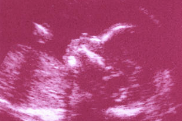
The sound waves hit tissues, body fluids, and bones. Waves then bounce back, like echoes, and are converted into pictures of the internal organs and—during pregnancy—the fetus.
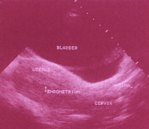
The images appear on a screen similar to a television. Dark areas show liquid, like amniotic fluid. Gray or light areas show denser material, like tissue or bone.
The type of ultrasound that is used most often combines still pictures to show movement, like the single frames that make a movie. This is called real-time ultrasound.
Uses of Ultrasound
Many people think of an ultrasound exam as an exam that is done during pregnancy. This often is true, but there are many other health conditions that can be monitored using ultrasound.
Obstetrics
Ultrasound is used in obstetrics to examine the growing fetus inside the mother’s uterus. A standard ultrasound can provide valuable information about the fetus’s health and well-being, including:
- Age of the fetus
- Rate of growth of the fetus
- Placement of the placenta
- Fetal position, movement, breathing, and heart rate
- Amount of amniotic fluid in the uterus
- Number of fetuses
- Some birth defects
If you have a high-risk pregnancy, your doctor suspects a problem, or your pregnancy continues after your due date, your doctor may want to use ultrasound to check on the well-being of your baby. Other uses of ultrasound during pregnancy include a fetal cardiogram. This is a detailed ultrasound exam of the heart that may be done if heart problems are suspected in the fetus. Ultrasound also may be used for diagnosing an ectopic pregnancy or determining a cause of bleeding or pain during pregnancy.
Gynecology
Ultrasound is used in gynecology to examine the pelvic organs. A ultrasound exam can help:
- Identify a pelvic mass
- Find causes of pelvic pain
- Find causes of abnormal bleeding or other menstrual problems
- Find the position of an intrauterine device (IUD)
- Diagnose and treat infertility
Types of Ultrasound
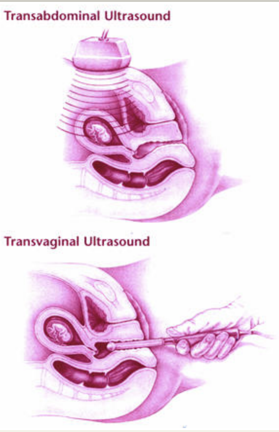
There are many different types of ultrasound exams. The type of ultrasound you have will depend on what types of images the doctor needs and why the exam is being done. The transducer may be placed on the abdomen (transabdominal) or in the vagina (transvaginal). Other types of ultrasound include:
- Doppler ultrasound: This test is performed the same way as transabdominal ultrasound. For this exam, higher-intensity sound waves are used to study the movement of blood (usually through the umbilical cord or between the uterus and placenta). It also can be used to listen to the baby’s heartbeat.
- Sonohysterography: For sonohysterography, you first will have a transvaginal ultrasound exam. A catheter (a small tube) then will be inserted through the cervix, and a saline solution (salt water) will be injected through the catheter. The saline solution fills the uterus so abnormal findings can be seen inside the uterus. It also acts as a contrast material, which makes it easier to see anything abnormal.
- Three- and four-dimensional (3D and 4D) ultrasound: These are types of transabdominal ultrasound. A 3D ultrasound exam takes thousands of images at once. These are stored and shaded to make a 3D image, which looks more lifelike. A 4D image is similar to a 3D image, but it also shows movement.
The Exam
An ultrasound exam may be done in a doctor’s office or a hospital. It may be performed by a doctor or a specially trained technician.
Transabdominal Ultrasound
If you are having a transabdominal ultrasound, wear loose-fitting clothes. This will allow your abdomen to be exposed easily. You may need to drink up to six glasses of water during the 2 hours before your exam. This will make your bladder full. A full bladder:
- Pushes loops of the bowel up and out of the way, making the uterus easier to see
- Moves the uterus higher in the belly, making the fetus easier to see
For this exam, you will lie on a table with your abdomen exposed from the lower part of the ribs to the hips. Mineral oil or a gel is applied to the surface of the abdomen. This improves contact of the transducer with the skin surface. Sound waves cannot move through air, so the gel helps get rid of air between the skin and the transducer. The hand-held transducer then is moved along the abdomen.
Transvaginal Ultrasound
For a transvaginal ultrasound, you will be asked to change into a hospital gown or undress from the waist down. You or your doctor may wish to have a chaperone present during the exam. You do not need to fill your bladder before the test. You will lie on your back with your feet in stirrups, like a pelvic exam. The transducer for this exam is shaped like a wand. It is covered with latex, like a condom, and lubricated before it is inserted into the vagina. This type of ultrasound can give a closer look at the pelvic organs and fetus.
Keepsake Ultrasounds
Today there are centers that use ultrasound to produce 3D videos and portraits of a fetus. These “keepsake” videos show facial features, fingers, toes, the baby’s sex, and movement.
Many parents enjoy having these moving portraits. However, not much is known about the effects of repeated exposure to ultrasound. It seems to be safe, but it is possible that problems could be found in the future.
Moreover, the workers at these centers often are not trained to interpret the images for you. Based on the ultrasound, you may be falsely reassured that your baby is doing well, when in fact there may be a problem. Or, you may be alarmed that the image shows an abnormality and, because these centers are not medical facilities, you will be left without any professional help. Obstetric ultrasound is best obtained through standard prenatal care.
After The Exam
After the test, your doctor will look at the images and discuss the results with you. He or she may consult with another doctor.
Finally…
Ultrasound is used to look for problems that may occur or to check on the progress of your pregnancy. It can be a useful tool, especially when used with other tests and exams. Your doctor will explain the reason for your ultrasound and tell you how to prepare for the exam.
Glossary
Amniotic fluid: Water in the sac surrounding the fetus in the mother’s uterus.
Cervix: The lower, narrow end of the uterus, which protrudes into the vagina.
Ectopic pregnancy: A pregnancy in which the fertilized egg begins to grow in a place other than inside the uterus, usually in the fallopian tubes.
Fetus: A baby growing in the woman’s uterus.
Intrauterine Device: A small device that is inserted and left inside the uterus to prevent pregnancy.
Placenta: Tissue that provides nourishment to and takes away waste from the fetus.
Transducer: A device that emits sound waves and translates the echoes into electrical signals.
Umbilical Cord: A cordlike structure containing blood vessels that connects the fetus to the placenta.
Uterus: A muscular organ located in the female pelvis that contains and nourishes the developing fetus during pregnancy.