Hysterosalpingography (HSG) is used to diagnose problems of the uterus and fallopian tubes. Most often it is used to see if a woman’s tubes are partly or fully blocked. It also can show if the inside of the uterus is of a normal size and shape. This page explains:
- reasons for HSG
- what to expect before and after HSG
- how it is done
- risks, complications, and alternative
Reasons for Hysterosalpingography
HSG is an X-ray procedure that is used to view the inside of the uterus and fallopian tubes. It checks for scarring or abnormalities in their size or shape, which can lead to infertility and pregnancy problems. HSG also is used a few months after some sterilization procedures to make sure that the fallopian tubes have been completely blocked.
What to Expect
HSG is done in a hospital, clinic, or health care provider’s office. It is best to have HSG done in the first half (days 1–14) of the menstrual cycle. This timing reduces the chance that you may be pregnant.
HSG is not done in a woman if she
- is pregnant
- has a pelvic infection
- has heavy uterine bleeding at the time of the procedure
Your health care provider may recommend that you take an over-the-counter pain reliever an hour before the procedure. Discuss this decision with your health care provider. In some cases, he or she also may prescribe an antibiotic for you to take before HSG. Most people can drive themselves home after having HSG. However, you may not feel well after the procedure, so you may want to make arrangements for someone to drive you home.
The Procedure
During HSG, a contrast medium is placed in the uterus and fallopian tubes. This is a fluid that contains a dye. The dye shows up in contrast to the body structures on an X-ray screen. The dye outlines their inner size and shape. It also is possible to see how the dye moves through the body structures.
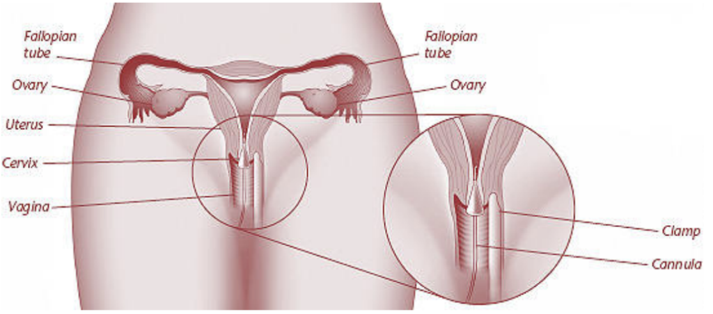
During hysterosalpingography, a dye is injected into the uterus through a plastic tube or cannula. X-ray images are taken as the dye moves through the uterus and into the fallopian tubes.
The procedure is performed as follows:
- You will be asked to lie on your back with your feet placed in stirrups as for a pelvic exam. A device called a speculum is inserted into the vagina. It holds the walls of the vagina apart to allow the cervix to be viewed. The cervix is cleaned.
- The end of the cervix may be injected with local anesthesia (pain relief). You may feel a slight pinch or tug as this is done.
- One of two methods may be used to insert the dye. In one method, the cervix is grasped with a device to hold it steady. An instrument called a cannula is then inserted into the cervix. In the other method, a thin plastic tube is passed into the cervical opening. The tube has a small balloon at the end that is inflated. The balloon keeps the tube in place in the uterus.
- The speculum is removed, and you are placed beneath an X-ray machine.
- The fluid slowly is placed through the cannula or tube into the uterus and fallopian tubes. The fluid may cause cramping. If the tubes are blocked, the fluid will cause them to stretch.
- X-ray images are made as the contrast medium fills the uterus and tubes. You may be asked to change position. If there is no blockage, the fluid will spill slowly out the far ends of the tubes. After it spills out, the fluid is absorbed by the body.
- After the images are made, the cannula or tube is removed.
After the Procedure
After HSG, you can expect to have a sticky vaginal discharge as some of the fluid drains out of the uterus. The fluid may be tinged with blood. A pad can be used for the vaginal discharge. Do not use a tampon. You also may have the following symptoms:
- Slight vaginal bleeding
- Cramps
- Feeling dizzy, faint, or sick to your stomach
Risks and Complications
Severe problems after an HSG are rare. They include an allergic reaction to the dye, injury to the uterus, or pelvic infection. Call your health care provider if you have any of these symptoms:
- Foul-smelling vaginal discharge
- Vomiting
- Fainting
- Severe abdominal pain or cramping
- Heavy vaginal bleeding
- Fever or chills
Alternatives
There are other procedures that can give your health care provider some of the same information as HSG:
- Laparoscopy—This surgical procedure requires general anesthesia.
- Hysteroscopy—This procedure can give a detailed view of the inside of the uterus. However, it cannot show whether the fallopian tubes are blocked.
- Sonohysterography—This technique uses ultrasound to show the inside of the uterus. Like hysteroscopy, it does not give information about the fallopian tubes.
Finally…
HSG is a way to diagnose problems of the uterus and fallopian tubes. The risks of HSG are low but you should know the warning signs of problems. Talk to your health care provider if you have questions about this procedure.
Glossary
Contrast Medium: A substance injected into the body that highlights internal structures during an imaging study.
Fallopian Tubes: Tubes through which an egg travels from the ovary to the uterus.
Hysteroscopy: A procedure in which a slender, light-transmitting device, the hysteroscope, is inserted into the uterus through the cervix to view the inside of the uterus or perform surgery.
General Anesthesia: The use of drugs that produce a sleep-like state to prevent pain during surgery.
Infertility: A condition in which a couple has been unable to get pregnant after 12 months of trying without the use of any form of birth control.
Laparoscopy: A surgical procedure in which a slender, light-transmitting instrument, the laparoscope, is inserted into the pelvic cavity through small incisions. The laparoscope is used to view the pelvic organs. Other instruments can be used to perform surgery.
Local Anesthesia: The use of drugs that prevent pain in a part of the body.
Sonohysterography: A procedure in which sterile fluid is injected into the uterus through the cervix while ultrasound images are taken of the inside of the uterus.
Sterilization: A permanent method of birth control.
Uterus: A muscular organ located in the female pelvis that contains and nourishes the developing fetus during pregnancy.
Source: acog.org